SUNDAY, 7 NOVEMBER 2010
 To a large extent, the behaviour of each cell in your body is determined by the environment that surrounds it. That environment influences whether a cell migrates, proliferates, becomes more specialised, or implements programmed cell death. The stiffness of the surfaces with which the cell interacts plays a key role in determining how a cell behaves. The biomechanical properties of cells are the focus of research being done by Ivan Minev, a second-year PhD student in the Department of Engineering.
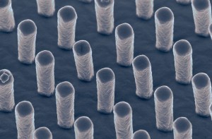
Ivan studies the responses of living cells to artificial surfaces. These surfaces consist of millions of rubber pillars, each with a diameter of approximately two micrometres (one micrometre is one thousandth of a millimetre).
Cells can exert small mechanical forces on the surfaces in contact with them; this means that cells sitting on Ivan’s artificial surfaces are able to deflect the rubber pillars. The amount of deflection gives the cells feedback about the stiffness of the surface, which in turn dictates how the cells behave. By altering the stiffness of the pillars, Ivan is able to study how the cells respond to different surfaces; he can also more accurately model how cells function within the body. For example, to study astrocytes (a cell type that mediates inflammation in the brain) it is much more appropriate to use surfaces with similar stiffness to those found in the brain. Ivan is able to control the stiffness of his surfaces by varying the heights and widths of the pillars, as well as by changing the material they are made from.

A process called photolithography is used to fabricate the surfaces. A mask consisting of transparent dots on an opaque background is placed over a layer of photosensitive epoxy resin that hardens upon exposure to light. Only the material beneath the transparent part of the mask is affected, creating columns of hardened material. A developing fluid is then used to wash away the unaffected material, leaving the pillars behind.
In the next step, the epoxy pillars are used as a template in a double casting procedure. Silicone polydimethylsiloxane, which is initially the consistency of honey, is poured onto the epoxy columns. After a night in an oven, the silicone becomes rubbery and is peeled away from the template. This creates a reverse mould, so that when the process is repeated, a silicone replica of the original pillars is produced.
This issue’s front cover shows a colour-enhanced scanning electron microscope image of one of these pillared surfaces, taken by Ivan and his colleague Rami Louca. A defect appears in the centre of the image, where the pillars have spontaneously collapsed onto one another. Different patterns of collapse can indicate that the sample has been accidentally scratched or that after immersing in fluid, the receding edges of drying droplets have caused the columns to be pulled over. The collapse does not tend to be a problem however, since one square centimetre is seeded with thousands of cells, and only a small proportion of the area has any defects.
In addition to helping researchers better understand cellular behaviour, artificial surfaces are potentially useful for a number of medical applications. For example, these surfaces could be used to minimise the ‘foreign body reaction’. An implant would be wrapped in an artificially soft surface, tailored to influence the response of immune cells, tricking them into accepting the implant as part of the body. Other possible applications include using the surfaces to promote nerve regeneration within a damaged spinal cord or to stimulate certain cells within the brain, in such a way that they help to alleviate the symptoms of Parkinson’s disease. If any of these endeavours are successful, artificial arrays of tiny pillars could lead to some novel medical materials and treatments.
Lindsey Nield is a PhD student in the Department of Physics
